Lotiskorea Newsletter Vol. 32
|
|
|
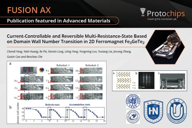
📖 In this newest research published in #AdvancedMaterials, researchers are looking at all-electric functional nanodevices with rapid reset and multi-state switching capabilities using our #FusionAX system!
💡Using an in situ biasing technique, and focus-ion beam preparation (FIB) the authors look at spin configuration rearrangement by domain wall number modulation in Fe3GeTe2.
⚡In-situ Lorentz transmission electron microscopy revealed the reduction of domain wall number corresponds to a decrease in resistance, new possibilities for manipulating magnetic states at the nanoscale are observed.
🖥️Additionally, simulations reveal the instant reversal of magnetic states to multi-domain wall configurations under single-pulse currents with higher amplitudes, driven by rapid thermal demagnetization.
🔬These groundbreaking results position 2D ferromagnets as intriguing candidates for the future of neuromorphic spintronics, offering unprecedented opportunities for innovation and discovery.
Want to read the entire research?
Find it here!
https://hubs.li/Q02DzMVk0
|
|
|
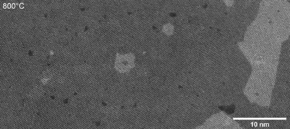
📖 With this newest published research atomic-scale mechanisms were observed on 2D materials using our #FusionAX system! The authors imaged, atom-by-atom, the thermally induced structural evolution of twisted bilayer transition metal dichalcogenides using in situ transmission electron microscopy.
🔥 The authors used short 0.5-s heat pulses from 100° to 1000°C and took STEM images in between to reduce thermal drift. It was observed at low temperatures that the moiré superlattices morphed into nanoscale aligned domains. It was observed that this process started by nucleating a new grain within one monolayer, its crystal orientation intricately templated by the other.
🔬Unexpectedly, the aligned domains grow through a mesmerizing dance of collective rotation of moiré supercells and the graceful hopping of 5|7 defect pairs at moiré boundaries. These observations offer unprecedented mechanistic insight into the atomic-scale interactions that govern moiré structures, illuminating the potential to pattern interfacial structure and properties of two-dimensional materials at the nanoscale.
📈These research results can be correlated to inconsistent and heterogeneous properties measured in moiré based devices. This can then lead to new synthesis, bottom-up routes, designing structures to control and pattern the twist angle on the nanoscale.
Want to read the entire research paper?
Find it here!
https://hubs.li/Q02tVHw50 |
|
|
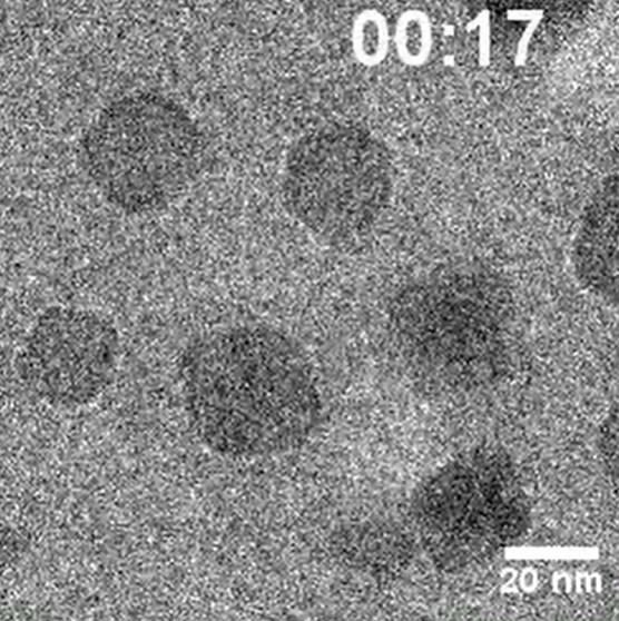
Core-shell (CS) nanoparticles are a promising candidate for catalysis. Due to lattice mismatch between the core and shell materials, there is an interfacial strain at the CS interface which extends into the bulk and this interfacial strain is responsible for the improved catalytic performance of the CS nanoparticles as compared to pure materials individually. In electrocatalytic applications, such as electrocatalytic water splitting, the catalyst undergoes redox kinetics and result in structural transformations which may impair the catalytic activity. Due to lack of sufficient spatiotemporal resolution in conventional characterization methods, it has been challenging to probe these structural transformations in the catalysts in real-time on individual nanoparticle level at atomic scale.
Researchers from Shanghai Jiao Tong University, UC Irvine and Irvine Materials Research Institute (IMRI) including Prof. Wenpei Gao and Prof. Xiaoqing Pan have used electrochemical liquid phase transmission electron microscopy (ECLPTEM) to study the structural dynamics of Pd@Pt CS nanoparticles under electrochemical (EC) cycling. They observed that etching and deposition of Pd involving migration of Pd atoms towards the surface followed by corrosion of Pt preferentially occurring at (111) terrace sites rather than edge or corner sites counterintuitively. The study provides insights to the structural evolution and surface reconstruction of CS electrocatalytic nanoparticles in real-time under EC cycling.
ECLPTEM experiments were performed on a JEOL 2800 microscope using a Protochips Poseidon liquid cell holder.
This in-situ phase-contrast time-series demonstrates the corrosion process in a octahedral Pd@Pt CS nanoparticles.
Read the interesting findings published in the journal Nature Communications.
https://lnkd.in/dCuJeZUP |
|
|
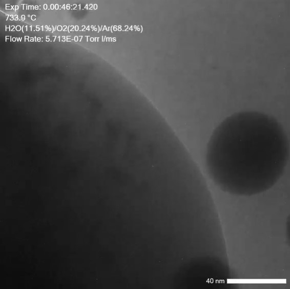
📖 In this newest publication in ACS Advanced Nano Materials, researchers from the #KULeuven have used the #AtmosphereAX system to look at passive adsorption materials for NOx removal. Authors Sreeprasanth Pulinthanathu Sree Claudio Bellani , Rainer Straubinger have investigated the dynamic behavior of palladium species impregnated onto zeolite under hydrothermal oxidation conditions, and using vapor!
🔬Using #Protochips' in situ gas phase TEM including the #AXON software, the formation and dispersion of Pd nanoparticles could be observed supported on a RHO zeolite. By heating the zeolite powder impregnated with a Pd complex up to 750°C within the TEM under an O2 atmosphere, water vapor, and argon, the dynamic evolution of Pd particles was shown.
🌡️ At lower temperatures, volatile masses began to form and gradually coalesced as the temperature increased. At the peak temperature of 750°C, a dispersal process occurred, releasing highly mobile smaller particles. This behavior underscores the intricate dynamics of Pd species under these conditions.
⚙️ The in situ TEM studies captured the dynamic changes of Pd nanoparticles, revealing mechanisms such as rapid thermally induced reduction and Ostwald ripening. These insights are crucial for optimizing and preparing supported noble-metal materials and catalysts.
Studies like these enhance the understanding of nanoparticle formation on supports for catalysis and underscore the potential of in situ TEM as a powerful method for investigating and improving the synthesis of various catalytic nanomaterials.
Read the entire paper here:
https://hubs.li/Q02zPSwJ0 |
|
|
📖 This latest research introduces a new methodology for synthesizing MoVSbOx catalysts for the ethane oxidative dehydrogenation (E-ODH) reaction using our #AtmosphereAX system. In this newest research it becomes clear that nanoscale and bulkscale can be used together to create better catalyst materials!
🔍💡With this new approach, M1 nanorods with unique facets were synthesized, with a large increase in the number of active sites. Through thermal activation it was shown that the combustion of amino-organic compounds generates defects along lateral facets, enhancing accessibility to additional active sites.
🔬 By doing in-situ electron microscopy analysis, the (trans)formation mechanism of multiple crystalline phases could be actively observed, shedding light on their role in catalytic activity. Plus, high-resolution HAADF-STEM revealed the location of active sites, providing invaluable insights into catalyst performance.
📈 Mo, V, and Sb elements together showed an increase in RedOx activity and selectivity during ethane conversion. Lastly, the catalysts in this research demonstrated high stability, maintaining impressive activity and selectivity over extended periods—a testament to their robustness and reliability.
⏰ In a continuous 480-hour experiment, conducted in a bench-scale reactor, our scaled-up quadrilobe-shaped catalyst showcased exceptional stability and performance under real-world conditions.
Want to find our more? Read the paper!
https://hubs.li/Q02v81f0 |
|
|
Are you interested in studying catalyst selectivity while observing corresponding structural changes? With #AtmosphereAX, you can visualize reactions at the nanoscale in real time while simultaneously analyzing gas streams, under relevant operating conditions. Interesting, right?
Here’s how this is uniquely possible with Atmosphere AX:
🛡️Preparation: The unique gas-handling manifold allows easy maintenance and cleaning, including a 50°C bake-out to remove carbon contamination! Additionally, with proactive pressure monitoring during experiments, the system automatically shuts down if a leak is detected, prioritizing the safety of the end-user and the microscope.
🔬Collection: During acquisition, AXON Synchronicity collects metadata from both the microscope and in situ system, allowing >400 pieces of metadata to be saved in each experiment, including critical parameters like heating ramp rates, pressure and gas composition.
💨Analysis: To accurately analyze selectivity, studying catalysts under relevant and realistic gaseous conditions is critical. Using volumetric blending, complex gas mixtures can be created and used with no need for calibration - you can even use and detect organic vapors such as benzene with ease!
👥Publishing: To support interdisciplinary research communities, #AXONStudio allows easy sharing of complete datasets, including corresponding metadata, with collaborators around the globe.
Using this workflow, you are well-equipped to scale your bulk catalysis experiments down to the nanoscale. This is done productively, reproducibly and in accordance with FAIR principles methods.
Want to know more about heterogeneous catalysis using in situ microscopy? Check out our blog: https://hubs.li/Q02CBb6c0
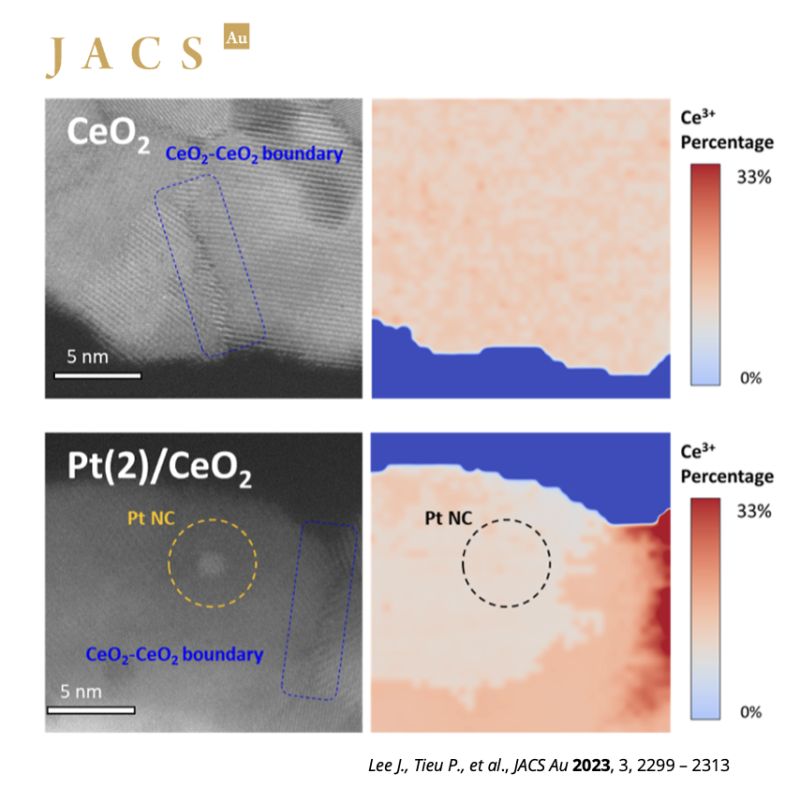
In the image, in situ STEM and EELS were used to investigate the influence of Pt single atoms and nanoclusters on the selectivity of H2 reactions on the surface of CeO2 catalysts. The study provided new insights into promoted CeO2 surface reactions with H2. For the full publication from UC Irvine and UC Santa Barbara, visit https://hubs.li/Q02CB93T0. |
|
|
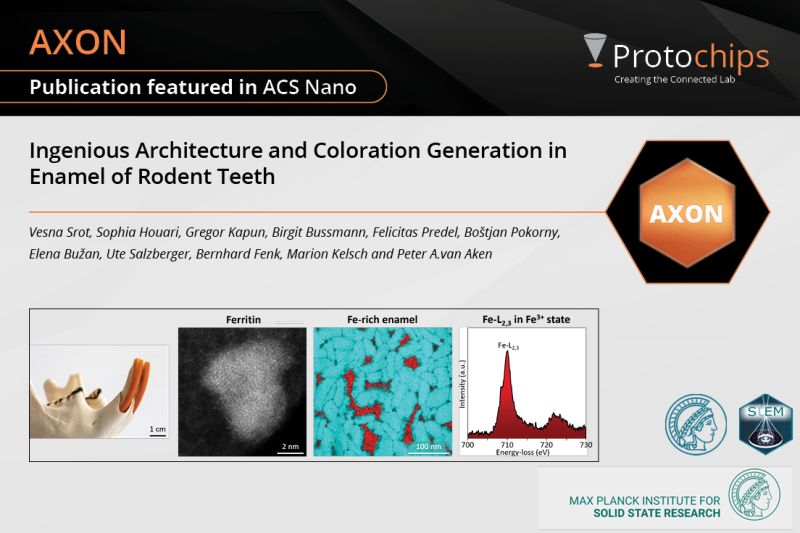
📖 In this, out-of-the-ordinary, post we want to focus on this newest work published in #ACSNano by Dr. Vesna Srot and Prof Peter A. van Aken! These authors have looked at continuously growing incisors in rodents and their microscopical structures. State-of-the-art direct atomic-scale imaging using our #AXONSynchronicity and #AXONDose software helped uncover the complex orchestration behind the formation of iron-rich enamel and surface layers in rodent incisors. 🦷
🎞️Through synergistic fusion of three-dimensional tomography and imaging techniques, these authors reveal a direct correlation between the presence of pockets infused with ferrihydrite-like material and the acid-resistant properties exhibited by the iron-rich enamel.
🖥️How was AXON Dose used? To overcome the electron beam sensitivity of the samples, low-dose (scanning) transmission electron microscopy (S)TEM was extremely important for this research! This is why AXON Dose was needed to control the electron dose well.
🔬Optical microscopy observations shed new light on the role of iron-rich enamel as a microstructural element that facilitates color transmission, challenging prevailing paradigms while maintaining the native color indistinguishable from regular enamel.
🚀 These discoveries reshape our understanding of incisor microstructure and color generation, with far-reaching implications for human health, the development of groundbreaking dental materials, and restorative dentistry.
Want to read the press release?
https://hubs.li/Q02BbVgB0
Want to read the entire paper?
https://hubs.li/Q02Bc6hT0 |
|
|
문의
Email : hskim@lotiskorea.com
Tel : 010-2858-2798 |
|
|
|